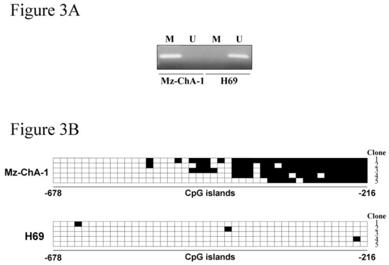
Figure 3. CpG island methylation within the socs-3 promoter is observed in Mz-ChA-1 but not H69 cells.

Genomic DNA extracted from the cell lines was treated with bisulfite and then subjected to methylation-specific PCR (A) using the methylated DNA- (M) and unmethylated DNA- (U) specific primer sets. PCR products were sequenced for the 44 CpG sites located between nucleotides -678 and -216 of the socs-3 promoter as described in the Materials and Methods section (B). The horizontal squares represent CpG islands while the vertical squares represent the individual 5 clones sequenced. Each black square represents a methylated cytosine residue within the CpG islands.
