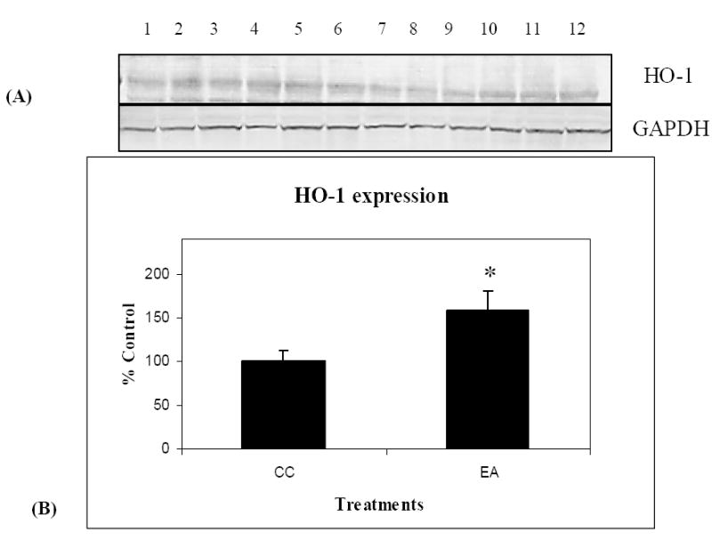
Figure 7.

HO-1 protein levels increase in response to treatment in aged canines. Western immunoblot analysis and quantification of canine brain homogenates samples containing 50μg of protein loaded onto 10% SDS-PAGE gels were completed using an anti-HO-1 antibody. A representative immunoblot (A) with lanes (1-6) representing the treatment group EA, and lanes 7-12 representing the control group CC is shown. GAPDH was used as a control for equal loading of protein. Densitometric values are plotted as a function of treatment group (B) showing a significant increase in HO-1 expression following the combined treatment of enriched environment and antioxidant-fortified food (EA) compared to control (CC). GAPDH densitometric data are represented as % control; ± SEM for each group. (n=6), * p <0.05.
