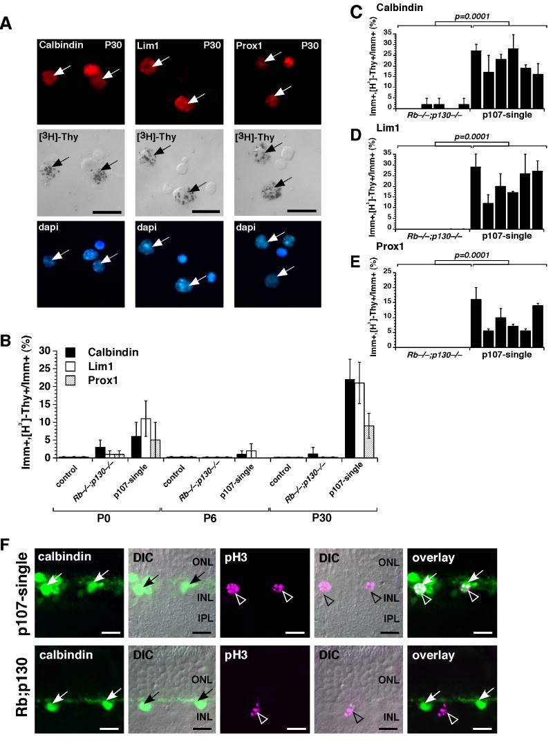
Figure 2. Horizontal cell proliferation in the postnatal mouse retina.
P30 p107-single retinae were [3H]-thymidine labeled for 1 hour and then dissociated, immunostained for Lim1, calbindin, and Prox1 expression and overlaid with autoradiographic emulsion to detect the [3H]-thymidine (A). The proportion of immunopositive cells that incorporated [3H]-thymidine was scored for each antibody at 3 stages of development (B). 6 animals from each genotype were assessed at each stage, and each sample was scored in duplicate. The controls were RbLox/Lox;p107+/−;p130−/− and Chx10-Cre;RbLox/Lox;p130−/− littermates. (C-E) Data on [3H]-thymidine labeling for individual animals at P30 is presented to illustrate the animal-to-animal variation with Chx10-Cre;RbLox/Lox;p130−/− littermates as a controls. (F) Co-immunolocalization of calbindin (green) and phospho-histone H3 (pH3) (purple) in P30 p107-single and control (Chx10-Cre;RbLox/Lox;p130−/−) littermates. Scale bars: 10 µm.

