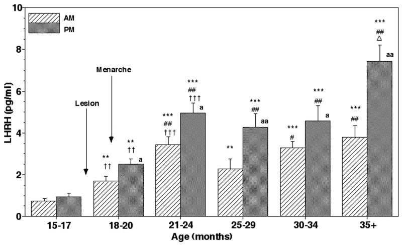
Figure 6.
Chronic effects of posterior hypothalamic lesions on LHRH release in the stalk-median eminence. Group means (± SEM) are shown. The number of lesion monkeys in each developmental period was 5–6. Approximate age of lesion and menarche are indicated by arrows. After lesions, both morning (AM) and evening (PM) LHRH levels were significantly higher than the pre-lesion values (within group) as well as the values in sham/lesion animals at the corresponding age (between groups). Note that LHRH levels further increased at 21–24 months of age, and stayed the same through 34 months of age. The evening levels after 35 months of age further increased over the levels at 21–24 months of age. The evening values were higher than the morning values in all examined periods after posterior hypothalamic lesions. ***: p<0.001 vs. 15–17 months (before lesion); **: p<0.01 vs. 15–17 months (before lesion); ## p<0.01 vs. 18–20 months; # p<0.05 vs. 18–20 months; Δ: p<0.05 vs. 21–24 months, 25–29 months, and 30–34 months; †††: p<0.001 vs. values of sham/controls at the corresponding age; ††: p<0.01 vs. values of sham/controls at the corresponding age; aa: p<0.01 vs. morning levels at each age; a: p<0.05 vs. morning levels at each age.

