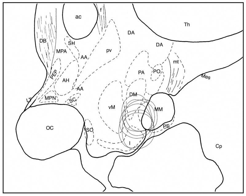
Figure 9.
Reconstruction of lesion area in the posterior hypothalamus of 6 monkeys shown in the sagittal plane. Schema was drawn based on Bleier’s atlas (2) with some modifications. Abbreviations: AA, Anterior hypothalamic area; ac, anterior commissura; AH, anterior hypothalamic nucleus; Cp, cerebral peduncle; DA, dorsal hypothalamic nucleus; DB, nucleus of diagonal bundle of Broca; DM, dorsomedial nucleus; f, fornix; I, infundibular nucleus; LT, lamina terminalis; Mes, mesencephalic tegmentum; MM, medial mammillary nucleus; MPA, medial preoptic area; MPN, medial preoptic nucleus; oc, optic chiasm; PA, posterior hypothalamic area; PE, perimammillary nucleus; PEP, periventricular preoptic nucleus; PM, paramammillary nucleus; PO, posterior nucleus; PV, paraventricular nucleus; SC, suprachiasmatic nucleus; SH, septohypothalamic nucleus; SO, supraoptic nucleus; Th, thalamus; VM, ventromedial nucleus.

