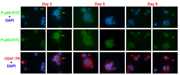
Figure 6.
Immunofluorescence microscopy analysis of phospho-Ser536-p65. Human mobilized peripheral blood CD34+ cells were cultured in Tpo and harvested on the indicated day of culture to analyze the expression and localization of phosphorylated p65 (Ser536) via immuno-fluorescence microscopy. Cells were co-stained with DAPI (blue) and antibodies against phosphor-Ser536-p65 (green) and CD41a (red). All images were captured using a 63X oil immersion objective.

