
Figure 1.
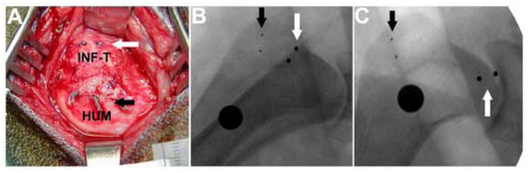
(A) To objectively assess repair connectivity, four small tantalum beads were implanted into the shoulder complex at the time of tendon repair: two embedded in the humerus (HUM) at the location of the black arrows and two on the surface of the infraspinatus tendon (INF-T) visualized at the white arrows. (B) Intra-operative fluoroscopy at the time of repair shows the tendon and bone beads as well as the calibration sphere (large black circle, OD = 9.5mm), (C) Follow-up fluoroscopy at 5 days post-op, demonstrates anatomic failure of the repair and approximately 2 cm retraction of the tendon stump.