
Figure 4.
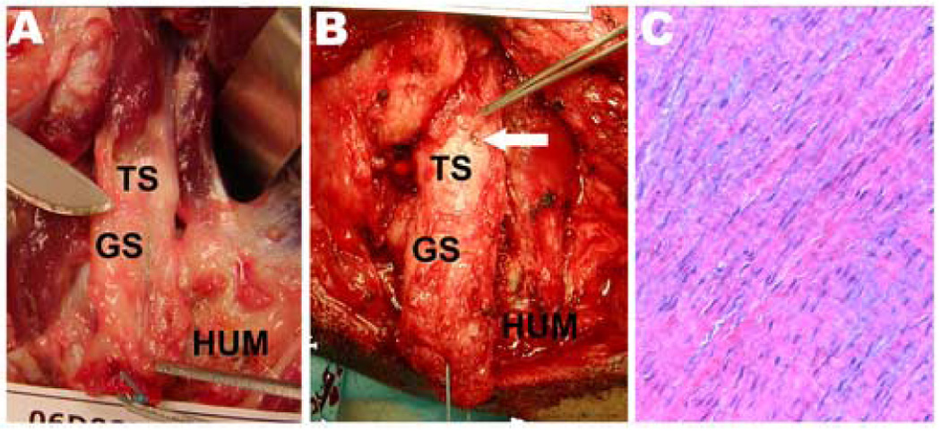
Robust, hypertrophic scar tissue formed in the 2–3 cm gap between the failed tendon stump (TS) and the humerus (HUM). Gap scar (GS) tissue at (A) three and (B) six weeks could be subjectively isolated as a tendon-like structure. The white arrow shows that one of the tantalum beads that had been placed on the tendon at surgery could be grossly visualized at dissection. (C) Histologically at six weeks, the isolated gap scar tissue demonstrates spindle-shaped nuclei and crimped, well-organized and oriented collagen (Hematoxylin-Eosin staining, 20X).