
Figure 4.
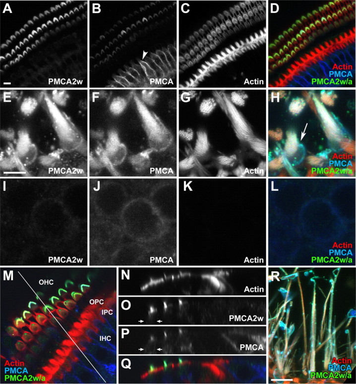
Localization of PMCA2w in rat hair bundles by immunofluorescence. A–D, Confocal cross sections through the P21 rat organ of Corti using R2w to detect PMCA2w (A; D, green), 5F10 to detect all PMCA isoforms (B; D, blue), and phalloidin to detect actin (C; D, red). Note the strong 5F10 immunoreactivity in basolateral membranes of inner hair cells (B, arrowhead). E–H, Utricular hair bundles were strongly labeled by R2w, as was the pericuticular necklace (H, arrow); the z-projection of two adjacent slices is shown. I–L, No R2w labeling of the lateral membranes of utricular hair cells; the z-projection of two adjacent slices is shown. M, En face view of a cochlea whole mount, with a line indicating the location of the x–z section for N–Q. OHC, Outer hair cells; OPC, outer pillar cells; IPC, inner pillar cells; IHC, inner hair cells. N–Q, The x–z section of cochlear whole mount shown in M. The color scheme in Q is the same as in M. PMCA2w immunoreactivity is strong in bundles of outer hair cells but absent from the cell bodies of outer hair cells (O, P, arrows), inner hair cells, and other cells of the organ of Corti. R, Lateral view of rat ampulla labeled with R2w. Scale bars: (in A) A–D, R, 10 μm; (in E) E–L, 5 μm; M, 91 × 91 μm; N–Q, 110 × 21 μm.