
Figure 6.
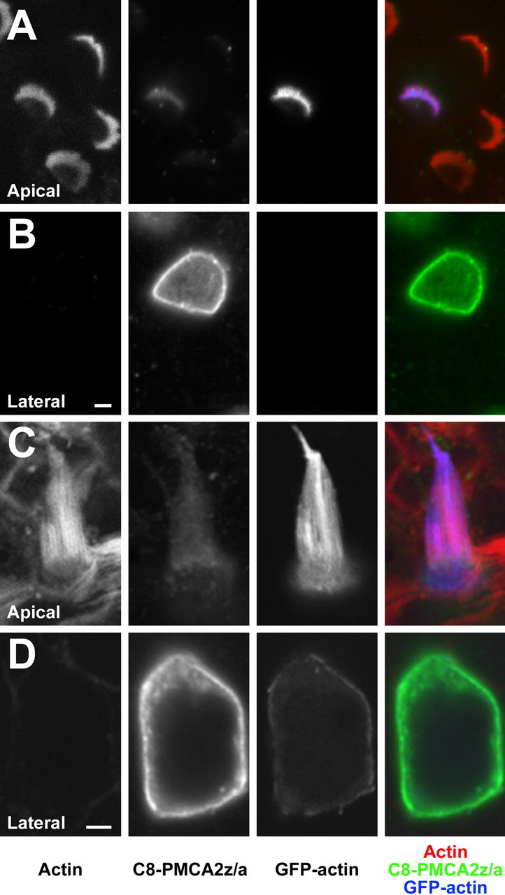
Targeting of PMCA2z/a in transfected rat hair cells. A, B, PMCA2z/a-C8 expression in the membrane of a cochlear outer hair cell at the level of the bundle (A) and near the nucleus (B). The magenta color of the hair bundle indicates lack of PMCA2z/a; the strong expression of PMCA2z/a on the basolateral membrane is apparent in B (green). C, D, PMCA2z/a-C8 on the membrane of a utricular hair cell, visualized at the level of the bundle (C) and near the nucleus (D). Green, Epitope-tagged PMCA2w/a; blue, GFP-actin; red, phalloidin to detect actin. Scale bars, 2 μm.