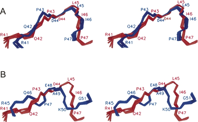
Figure 3.
Stereoview of the superimposed polar loop regions (residues 41–47) in the c32–52 peptide and in the full-length subunit c structures. (A) The c32–52 structure (red) and the NMR structure of the E. coli subunit c (1C0V, blue). The RMSD for backbone atoms is 1.1 Å. (B) The c32–52 structure (red) and the X-ray crystal structure of the subunit c from Ilyobacter tartaricus (1YCE, blue). The RMSD for backbone atoms is 2.4 Å.

