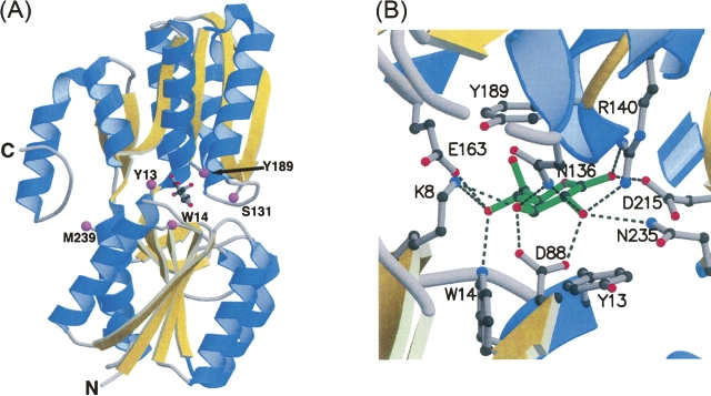
Figure 2.
Crystal structure of tmGBP. (A) The overall structure complexed with glucose (ball and stick) including sites of cysteine mutations for fluorophore attachment (magenta spheres). (B) Close-up view of the binding pocket: (green: glucose; dashed line: hydrogen bonds). The figure was generated using MOLSCRIPT (Kraulis 1991) and RASTER3D (Merritt and Murphy1994).

