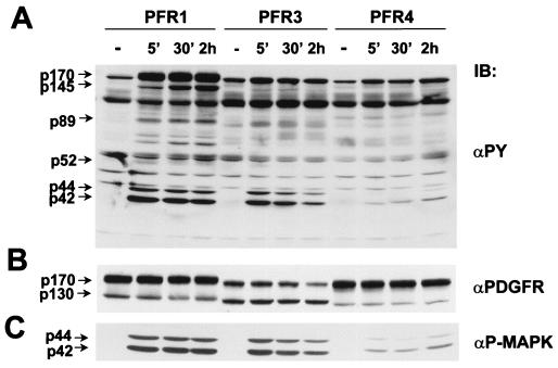
Figure 3.
Time course of induced protein tyrosine phosphorylation of PC12 cell lines expressing PFR1, -3, and -4. Cells were untreated (−) or incubated at 37°C with PDGF (30 ng/ml) as indicated. (A) Total cell extracts (50 μg) were immunoblotted with an anti-phosphotyrosine (αPY). Molecular mass standards (kDa) are shown. (B) The same blot was stripped and the top part reprobed with anti-PDGFR. The two forms of the chimeras are indicated. (C) The bottom part of the gel in A was reprobed with anti-active MAPK. p44ERK-1 and p42ERK-2 are indicated.

