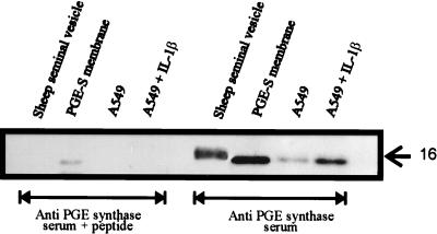
Figure 8.
Western blot analysis of PGE synthase in A549 cells and sheep seminal vesicles. The microsomal fractions (10 μg) from cells grown for 24 h in the presence (1 ng/ml) or absence of IL-1β were fractionated by SDS/PAGE and were transferred to poly(vinylidene difluoride) membrane. The membranes were incubated by using either PGE synthase antiserum or PGE synthase antiserum containing the antigenic peptide (10−6 M). Also analyzed was the commercially available, partly purified PGE synthase from sheep seminal vesicles (6 μg) as well as the membrane fraction from bacteria expressing human PGE synthase (1 μg).

