
Fig. 2. SgI localization is low in the drusen of AMD donors.
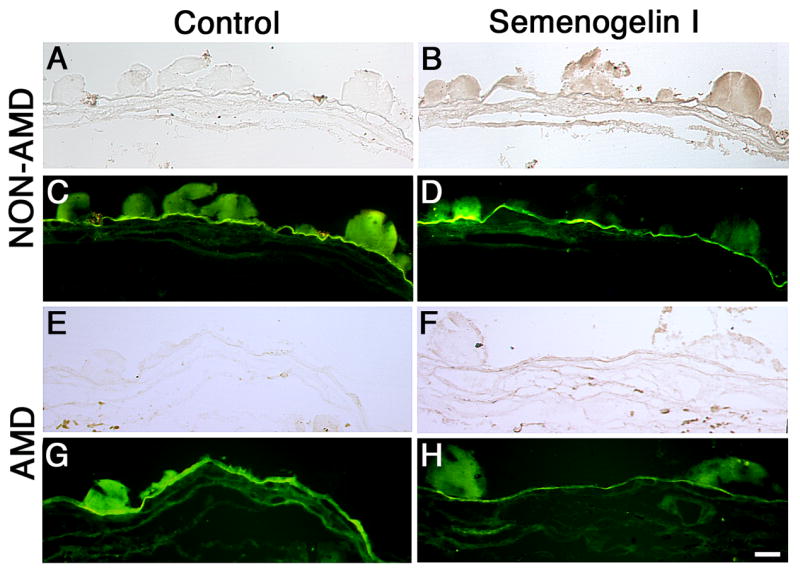
SgI staining is significantly reduced in the drusen from the AMD donors. 5μm paraffin sections of isolated Bruch’s membrane and choroid from a human donor previously diagnosed with AMD (E–H) and non-AMD eyes (A–D) were probed with SgI antibody in 5% BSA, PBS and 0.3% Triton-X100 overnight at 4°C. Sections were washed, incubated with secondary antibody, conjugated to biotin for 1h at RT, washed, and incubated with avidin in PBS for 30 min, then developed with DAB for 2 minutes. The controls (A, C, E, G) were omitted the antibodies and did not display any labeling. The sections were examined in bright-field (A, B, E, F) or FITC channel (C, D, G, H). Observation of the samples in the FITC channel revealed the autofluorescence of the Bruch’s membrane and drusen. 5 control and 8 AMD donors were analyzed. Bar = 200μm.