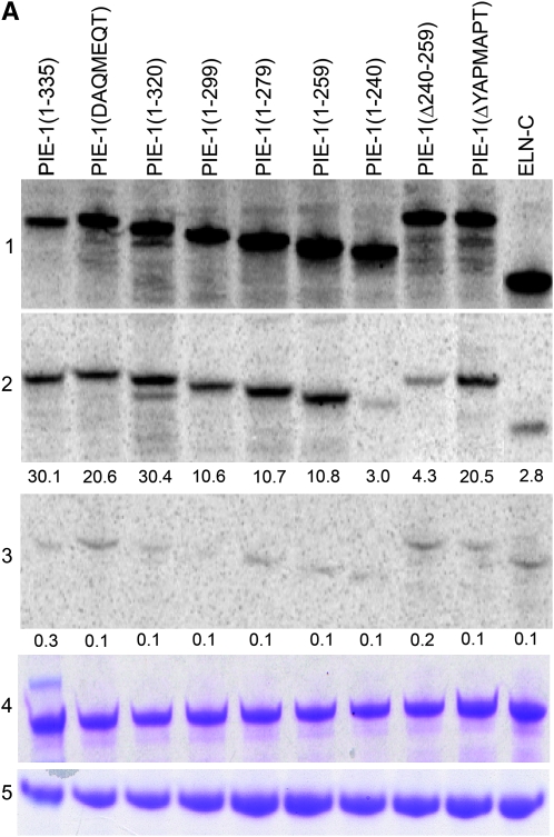
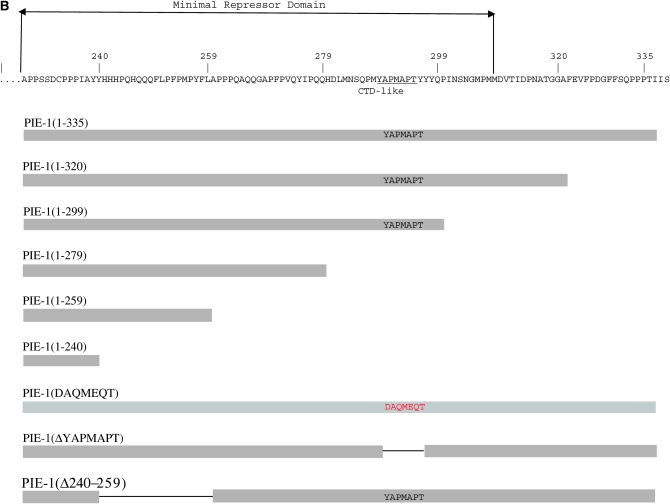
Figure 1.—
PIE-1 binds to CIT-1.1 in vitro. (A) In vitro translated and 35S-labeled full-length PIE-1 and mutant derivatives (section 1—input) were incubated with immobilized MBP:CIT-1.1 (section 2) or negative control MBP:PAR-5 (section 3) and bound proteins were resolved by SDS–PAGE. Sections 4 and 5 show Coomassie staining of MBP:CIT-1.1 (section 4) and MBP:PAR-5 (section 5) to control for loading. ELN-C is elongin C (DeRenzo et al. 2003) used here as a negative control. Numbers below sections 2 and 3 indicate percentage bound (bound/input × 100%), as calculated by measuring band intensities using Imagequant software (Molecular Dynamics). (B) Diagram showing the sequence of the C-terminal domain of PIE-1 and the mutant derivatives used in this study. The minimal repressor domain is the minimal PIE-1 fragment that can inhibit transcription when artificially brought to a promoter in HeLa cells (Batchelder et al. 1999).


