Figure 6.
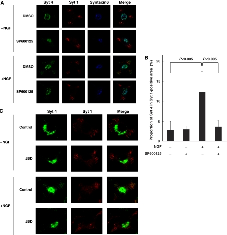
Requirement of JNK for NGF-dependent localization of Syt 4 to mature dense-core vesicles in PC12 cells. (A, B) PC12 cells were preincubated with 20 μM SP600125 (+) or DMSO (−) for 30 min, followed by a further 4 h incubation in the presence or absence of 100 ng/ml NGF. (C) PC12 cells were infected with retrovirus encoding JBD, or with corresponding control retrovirus (control). Infected cells were incubated for 4 h in the presence or absence of 100 ng/ml NGF. (A, C) The cells were fixed and subjected to immunocytochemistry with antibodies to Syt 4, Syt 1 (a marker for mature dense-core vesicle) and Syntaxin6 (a marker for Golgi body and immature secretory vesicle). Fluorescence images were recorded by a confocal laser-scanning microscope and typical images of cells are shown. (B) Quantification of the intracellular distribution of Syt 4. The cells were fixed and subjected to immunocytochemistry with antibodies to Syt 4, and Syt 1. The integrated fluorescence intensities of Syt 4 within the whole cell (a) and of Syt 1-positive (b) areas were detected by a confocal laser-scanning microscope. The proportion of (a) to (b) was calculated from 20 independent images by MetaMorph software. Data are means±s.d. P<0.005, Student's t-test.

