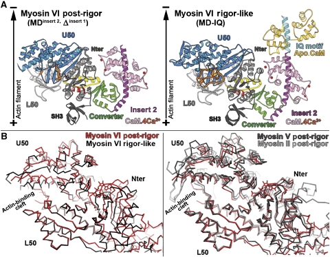
Figure 2.
The post-rigor state of myosin VI. (A) The myosin VI post-rigor structure is compared to that in the rigor-like state (right). The motors are oriented as if they are bound to a vertical actin filament (black arrow). Note the position of the lever arm, which is very similar in the rigor-like and post-rigor states. (B) On the left, porcine myosin VI in the post-rigor state (red) is compared with the myosin VI rigor-like state (black) after superposition of the L50 subdomains. Note the difference in the position of the Nter and U50 subdomains. The actin-binding cleft is more open in the post-rigor state (red) compared with the rigor-like state (black). On the right, three different post-rigor state structures are superimposed (chicken myosin V in black, Dictyostelium myosin II in gray and porcine myosin VI in red). Note that the positions of their subdomains are quite similar.

