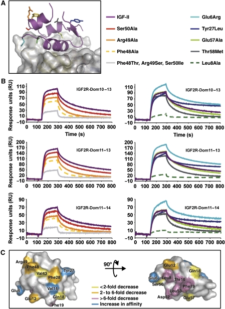
Figure 4.
Novel IGF-II mutations. (A) A view of the IGF-II/IGF2R-Dom11–13 complex with side chains investigated by mutagenesis shown as sticks and coloured according to the legend. (B) BIAcore analyses of IGF-II and analogues (representative curves each at 50 nM) binding to multi-domain IGF2R fragments. Curves are coloured as in the legend to panel A. (C) Summary of all IGF-II mutagenesis studies, which used IGF2R fragments. IGF-II side chains are coloured according to their effect on IGF2R binding (see legend).

