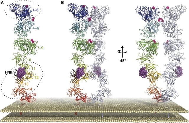
Figure 6.
Putative model for the relative arrangement of IGF2R extracellular domains. (A) View of an IGF2R monomer, coloured from blue at the N-terminus to red at the C-terminus, with FNII in black, IGF-II in magenta and mannose-6-phosphate-binding sites indicated by pink spheres. Dotted ellipses indicate regions of the model for which X-ray crystallography structures have been solved (bovine domains 1–3 and human domains 11–14). (B) Views of a tentative IGF2R dimer, based primarily on crystal packing observations for domains 11–14 and domains 11–13. For each dimer, one monomer is coloured as in panel A and the other is coloured gray/blue.

