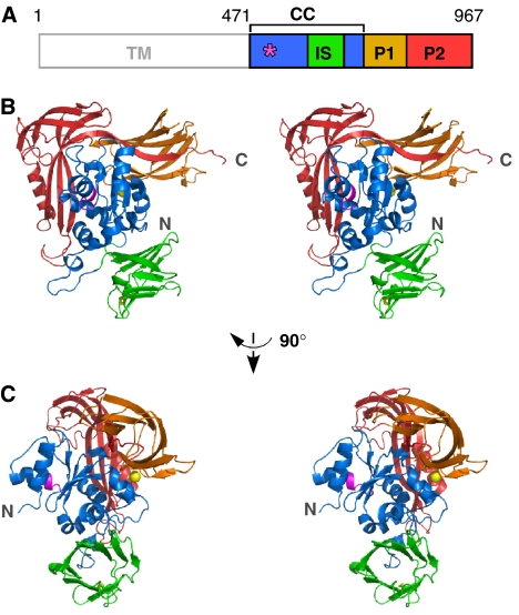
Figure 3.
Crystal structure of the C-terminal soluble domain of STT3. (A) Domain structure of P. furiosus STT3. TM, transmembrane domain; CC, central core domain, residues 471–600+683–725; IS, insertion domain, residues 601–682; P1, peripheral domain 1, residues 726–821; P2, peripheral domain 2, residues 822–967. The position of the WWDYG motif is indicated by an asterisk. (B) Stereoview of the overall structure of the C-terminal soluble domain of STT3 (residues 471–967). The WWDYG motif is shown in magenta. The disulfide bond between C638 and C658 is shown as yellow sticks. A bound metal cation is shown as a yellow sphere. (C) Different view from (B).

