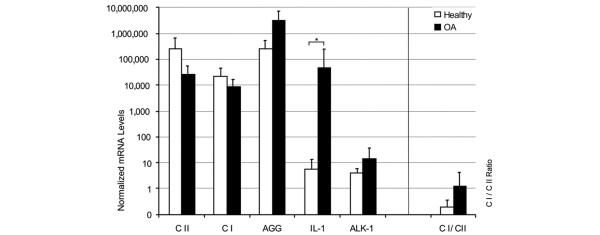
Figure 1.
Gene expression patterns of chondrocytes ex vivo. Chondrocytes were isolated from cartilage of healthy individuals (n = 6, white bars) or osteoarthritis (OA) patients (n = 20, black bars). The ex vivo gene expression of type I and II collagen (CI and CII, respectively), aggrecan (AGG), IL-1β and activin-like kinase (ALK)-1 was determined using qRT-PCR. The mRNA levels were normalized to GAPDH and amplified by a factor of 106. The collagen type I to collagen type II mRNA ratio was calculated as a measure for the differentiation status of the chondrocytes. Statistically significant differences (p < 0.05) are marked by asterisks (*).

