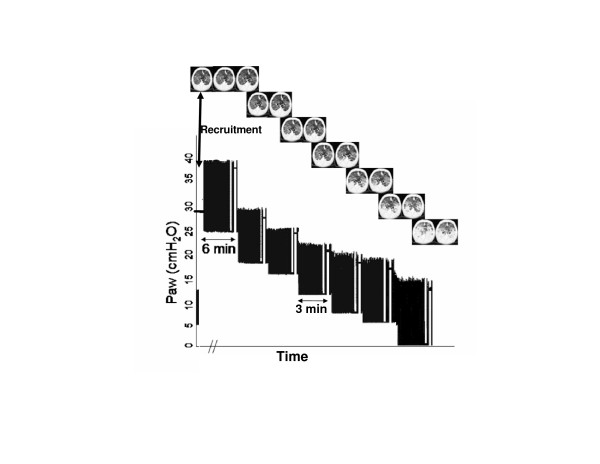
Figure 1.
Time plot of Paw during the PEEP titration procedure. The baseline ventilation, with a PEEP of 5 cmH2O, and the recruitment maneuver followed by the descending PEEP titration are shown. At the end of each PEEP step, a CT scan was performed at end-expiratory (left) and end-inspiratory (right) pauses. (CT scan images from a representative animal are shown.) CT, computed tomography; Paw, airway opening pressure; PEEP, positive end-expiratory pressure.

