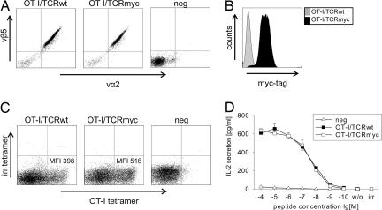
Fig. 2.
OT-I/TCRmyc is expressed and functions comparably to OT-I/TCRwt. (A) The TCR-deficient 58 cells were transduced with the indicated OT-I TCR constructs. Cells were enriched with vβ-chain-specific antibodies and analyzed by flow cytometry with vα2- and vβ5-specific antibodies. Untransduced cells (neg) served as a negative control. (B) The 58 cells transduced with the OT-I/TCRmyc were stained with an antibody specific for the myc-tag sequence. Cells transduced with the wild-type receptor were used as a control. (C) The B6 splenocytes were transduced with either OT-I/TCRwt or OT-I/TCRmyc and after 72 h were stained with a CD8-specific antibody, an OT-I specific MHC-tetramer, and an irrelevant tetramer (irr). Cells are gated on CD8 expression. Numbers indicate the MFI of the tetramer staining. (D) The 1 × 10⁵ OT-I/TCRwt- or OT-I/TCRmyc-transduced 58 (CD8α+) cells or untransduced cells (neg) were stimulated for 24 h with 1 × 10⁵ T2-Kb cells pulsed with ova peptide. IL-2 concentration of the culture supernatant was analyzed by ELISA. Unloaded T2-Kb cells (w/o) or T2-Kb cells loaded with irrelevant peptide (irr) served as negative controls. Data represent the mean values of duplicates, and error bars indicate SD.

