Fig. 5.
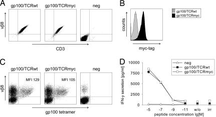
The human gp100/TCRmyc is expressed and functions comparably to gp100/TCRwt. (A) The human T cell line Jurkat76 was transduced with gp100/TCRwt or gp100/TCRmyc, enriched with vβ8-chain specific antibodies, and subcloned. TCR expression was analyzed by flow-cytometry staining with a vβ8-specific antibody. Untransduced cells (neg) served as a negative control. (B) Jurkat76 cells transduced with gp100/TCRmyc were stained with a myc-specific antibody and analyzed by flow cytometry. Cells transduced with the wild-type receptor served as a control. (C) PBLs were transduced with gp100/TCRwt or gp100/TCRmyc vectors and were stained with a vβ8-specific antibody and a gp100-specific tetramer. Untransduced PBLs (neg) show the endogenous vβ8-positive T cells. Numbers indicate the MFI of the tetramer staining. (D) gp100/TCRwt- or gp100/TCRmyc-transduced PBLs were cocultured with T2 cells pulsed with gp100 peptide for 24 h. Untransduced PBLs were used as a negative control (neg). Culture supernatant was analyzed for IFN-γ concentration by ELISA. Unloaded T2 cells (w/o) or T2 cells loaded with irrelevant peptide (irr) served as negative controls. The data represent mean values of duplicates, and error bars indicate SD. The results were reproduced in two independent experiments and with two different donors.

