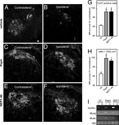
Fig. 4.
Pep4 and NEP1–40 prevent motor neuron death after sciatic nerve axotomy in neonate mice. (A–F) Photomicrographs showing ChAT immunoreactivity in the lumbar spinal cord of neonate mice. The sciatic nerve was axotomized and animals were treated during 5 days with vehicle (A and B), 11.6 mg/kg body mass Pep4 (C and D), or NEP1–40 (E and F). The sides contralateral (A, C, and E) and ipsilateral (B, D, and F) to the lesion are shown. (Scale bar, 25 μm.) (G and H) Quantification of the numbers of surviving motor neurons as shown in A–F after ChAT immunoreactivity (G) and toluidine blue staining (H). Data are presented as percentages of motor neurons in the corresponding side contralateral to the lesion [*, P < 0.05 vs. vehicle (Vh); n = 9–11 mice per group]. (I) Representative RT-PCR showing the mRNA levels of nicotinic acetylcholine receptor α-subunit (AchRα), myogenin (Myog), and muscle-specific kinase (Musk) in the hindlimb muscles contralateral (C) and ipsilateral (I) to the lesion. 18S rRNA levels are shown as internal control.

