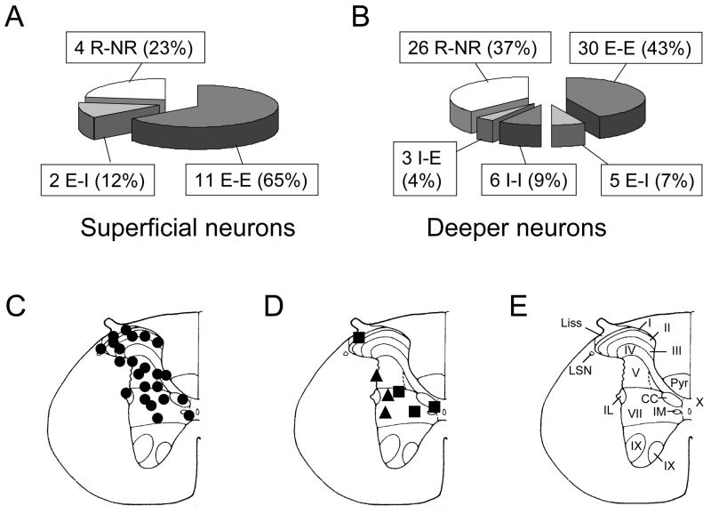
Fig. 1.
Recording sites and response patterns of low thoracic (T9-T10) spinal neurons to gastric distension (GD) and duodenal distension (DD). A, B: Comparison of superficial and deeper spinal gastroduodenal convergent neurons. E, excitatory response. I, inhibitory response. R, response. NR, no response. First response is to GD. Second response is to DD. C: Locations of spinal neurons responding to both GD and DD. The black circles represent neurons excited by both GD and DD. D: The black squares represent neurons excited by GD but inhibited by DD. The triangles represent neurons inhibited by GD but excited by DD. E: Schematic drawing of the T10 spinal segment (Molander et al. 1984). I-X indicates laminae; Liss, Liss’s tract; LSN, lateral spinal nucleus; Pyr, pyramidal tract; IL, intermediolateral nucleus. IM, intermediomedial nucleus. CC, column of Clarke.

