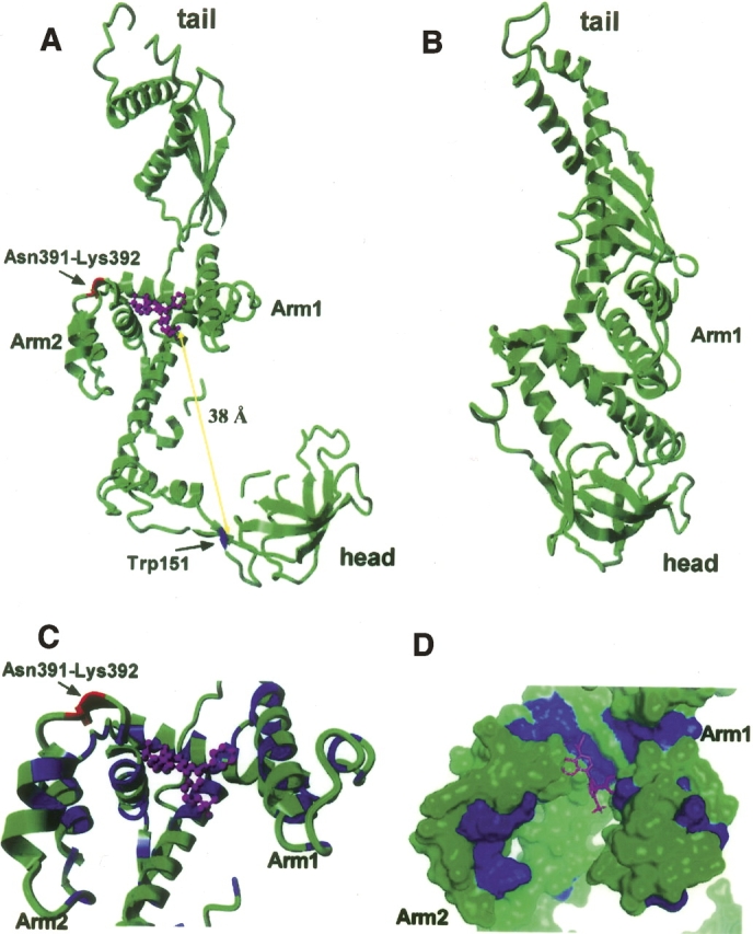
Figure 8.

Docking of bis-ANS into the TF crystal structure. (A) TF PDB data 1w26 (Ferbitz et al. 2004) and the programs Hex 4.5 and Yasara were used to dock bis-ANS into the TF crystal structure. bis-ANS is shown in magenta, Asn391-Lys392 in red, and Trp151 in blue. The distance between Trp151 and the putative bis-ANS binding site is consistent with the observation of excitation energy transfer (Fig. 2A). (B) Crystal structure of TF378 (Ludlam et al. 2004), in which no docking site for bis-ANS could be found. (C) Details of the bis-ANS binding region; bis-ANS is shown in magenta, Asn391-Lys392 in red, hydrophobic residues are blue, and hydrophilic residues are green. (D) Molecular surface of TF; bis-ANS is shown in magenta, hydrophobic residues are shown in blue, and hydrophilic residues in green.
