Figure 2.

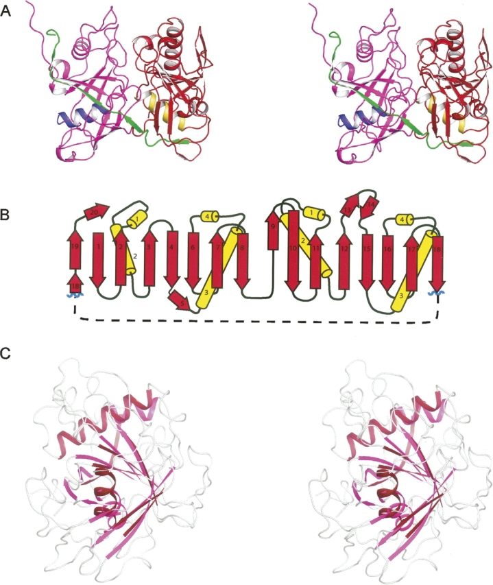
Structural representations and topology of apo PrpF. (A) A stereo ribbon representation of monomer A where the N- and C-terminal domains are colored in magenta and red, respectively. The central α-helix of the N- and C-terminal domains are depicted in blue and yellow, respectively, whereas the final β-strands that extend across both domains are colored in green. (B) The topology of the two domains. (C) A stereo superposition of the N- and C-terminal domains where the structurally similar secondary structural elements of the barrel are depicted in magenta and red for the N- and C-terminal domains, respectively. Figures 2–5 were prepared with the program PyMol (DeLano 2002).
