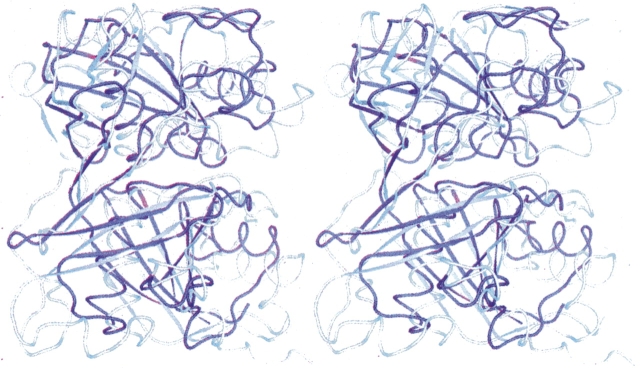
Figure 4.
Stereo overlay of apo PrpF and diaminopimelate epimerase (2GKJ). The β-strands for PrpF are depicted in light blue, whereas the helices and random coil are shown in white. In contrast, the entire molecule for diaminopimelate epimerase is colored in dark blue. The superposition was performed with the program ALIGN (Cohen 1997).

