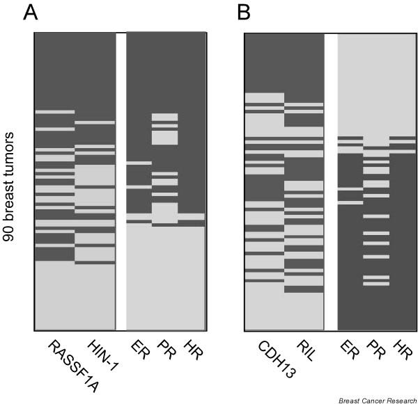
Figure 3.
Comparison of two methylation panels with hormone receptor status. (a) Dichotomous heat map representing DNA methylation in the HIN-1/RASSF1A panel (left) and hormone status of each tumor (right). (b) Dichotomous heat map representing DNA methylation data in the RIL/CDH13 panel (left) and hormone receptor status of each tumor (right). Dark gray, positive; light gray, negative.

