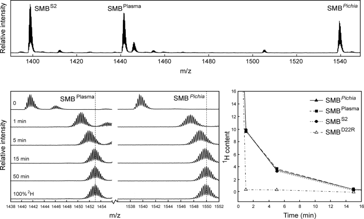
Figure 2.
Comparison of global hydrogen (1H/2H) exchange properties of different preparations of SMB. (Upper panel) A full scan mass spectrum of the quadruple charge state of the three forms of the SMB domain that we tested simultaneously in this study. The monoisotopic masses are shown, which translate into the following neutral molecular masses—SMBS2: 5588.33 Da (Δm = 0.17 Da); SMBplasma: 5758.51 Da (Δm = 0.18 Da); and SMBPichia: 6150.67 Da (Δm = 0.21 Da). Deviations from the theoretical masses are shown in brackets. SMBplasma contains an additional molecular species (5776.50 Da) due to an incomplete conversion between homoserine and homoserine lactone after CNBr cleavage. (Lower left panel) The mass spectra obtained for SMBplasma and SMBPichia are shown as a function of exchange time after dilution into deuterium oxide at 25°C. Notably, only one isotope envelope is produced by this exchange experiment for each SMB variant. (Lower right panel) The residual 1H content as a function of exchange time is illustrated for the three different wild-type SMB preparations, as well as a single-site mutant (SMBD22R) with impaired binding to PAI-1 and uPAR.

