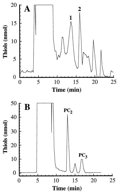
Figure 7.
AtPCS1-FLAG-dependent PC synthesis in vivo and in vitro. (A) Reverse-phase FPLC analysis of nonprotein thiols in the soluble fractions extracted from DTY167/pYES3-AtPCS1∷FLAG cells after growth in liquid medium containing CdCl2 (50 μM). Peaks 1 and 2 were found to comigrate with PC2 and PC3 standards, respectively, purified from S. pombe. The equivalent fractions extracted from DTY167/pYES3-AtPCS1∷FLAG cells after growth in medium lacking CdCl2 and from DTY167/pYES3 cells after growth in medium containing CdCl2 (50 μM) were devoid of PC-like nonprotein thiols. (B) Reverse-phase FPLC analysis of the nonprotein thiols formed after incubation of GSH (3.3 mM) with immunoaffinity-purified AtPCS1-FLAG in the presence of Cd2+ (200 μM). The peaks designated “PC2 ” and “PC3 ” were identified on the basis of their Glu/Gly ratios (2.1 ± 0.1 and 2.9 ± 0.2, respectively) and comigration with S. pombe PC standards.

