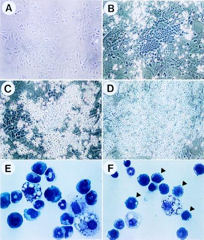
Figure 1.
Characterization of hematopoietic cells generated in the co-culture. Fetal liver-derived HSCs (E14.5) were cultured on primary fetal hepatic cells for 10 days in the presence (B, C, and E) or absence of Dex (D and F). (A) Morphology of the fetal hepatic culture (E14.5) just before the addition of HSCs. (B) Typical cobblestone area found in the co-culture. (C and D) Two different floating cell types found in the co-culture. (E and F) Cytospin preparations of cells from the Dex-plus (E) or Dex-free (F) culture. Arrowheads indicate the cells specifically appeared in the Dex-free culture.

