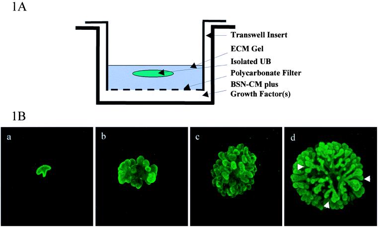
Figure 1.
A novel system for in vitro branching morphogenesis of the UB. (A) The culture system. UBs free from mesenchyme were microdissected from E-13 rat kidney rudiments and placed in an ECM gel suspension composed of type I collagen and growth factor-reduced Matrigel and cultured in BSN-CM supplemented with 10% FCS and growth factors. Details are given elsewhere in the text. The cultured UB was monitored daily by microscopy. (B) The UB undergoes branching morphogenesis in vitro and develops three-dimensional tubular structures in the absence of mesenchyme. E-13 rat UB was isolated and cultured as described. After culture, UBs were fixed at different time points and processed for DB lectin staining. Three-dimensional reconstructions of confocal images are shown. (a) A freshly isolated UB from an E-13 rat embryonic kidney with a single, branched tubular structure. (b) The same UB shown in (a) cultured for 3 days. The tissue has proliferated and small protrusions have formed. (c) The same UB shown in (a) cultured for 6 days. More protrusions have formed, and the protrusions have started to elongate and branch dichotomously. (d) The same UB shown in (a) cultured for 12 days. The protrusions have undergone further elongation and repeated dichotomous branching to form a structure resembling the developing collecting system of the kidney. The white arrows indicate branch points. At higher power, the structures formed in this in vitro culture system exhibited lumens. Phase microscopic examination and staining for markers revealed no evidence for contamination by other tissue or cells.

