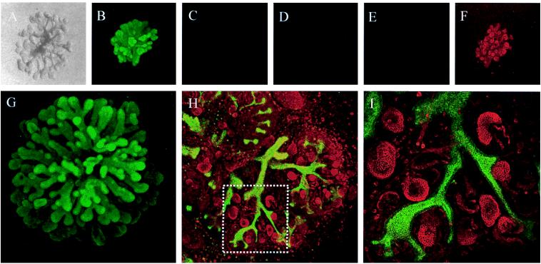
Figure 6.
The cultured three-dimensional tubular structure exhibits markers of UB epithelium and is functionally capable of inducing nephrogenesis when recombined with metanephric mesenchyme in vitro. (A– F) The cultured three-dimensional tubular structure exhibits markers of UB epithelium. The UBs were cultured in the presence of BSN-CM and GDNF and then stained for various markers. (A) Light microscopic-phase photograph of cultured UB. (B) Staining with DB lectin, a ureteric bud-specific lectin that binds to the UB and its derivatives. (C) Staining for vimentin, a mesenchymal marker. (D) Staining for neural cell adhesion molecule, the early marker for mesenchymal-to-epithelial conversion in the kidney. (E) Staining with PNA lectin, a mesenchymally derived renal epithelial cell marker. (F) Staining for cytokeratin, an epithelial marker. (G–I) The cultured three-dimensional tubular structure is capable of inducing nephrogenesis when recombined with metanephric mesenchyme. The isolated UB was first cultured 7–10 days as shown in G. Then, the cultured UB was removed from the ECM gel and recombined with freshly isolated metanephric mesenchyme from E-13 rat kidneys. The recombinant was cultured on a Transwell filter for another 5 days. After culture, the sample was double-stained with DB lectin (FITC) and PNA lectin (tetramethylrhodamine B isothiocyanate) as shown in H and in the enlarged section of H shown in I. Results indicate that the in vitro cultured UB-derived structures are capable of inducing nephrogenesis in vitro.

