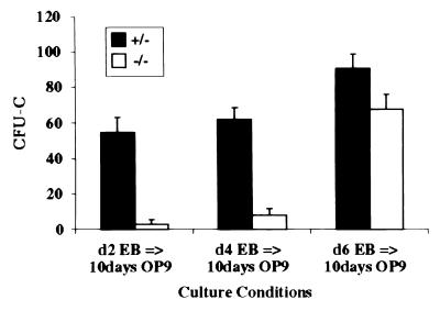
Figure 3.
A time course of myeloid–erythroid colony assays derived from switch cultures. ES cells differentiated as EBs for 2, 4, or 6 days, then were passaged onto OP9 stromal cells for 5 days, then replated onto OP9 cells for an additional 5 days. Colony assays were performed on the day 10 OP9 cultures. The number of CFU-Cs (the y-axis) is calculated per 5 × 10⁵ day 10 OP9 input cells. The black and white bars represent flk-1 (+/−) and flk-1 (−/−) cultures, respectively. No obvious differences in colony types were apparent between flk-1 (+/−) and flk-1 (−/−) cultures.

