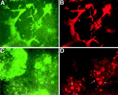
Figure 5.
β-gal and CD31 expression in flk-1 (+/−) and flk-1 (−/−) EBs. Photomicrographs of day 8 after dispase attached EB cultures stained for β-gal activity with FDG (A and C) and CD31 expression stained with phycoerythrin-conjugated anti-CD31 (B and D). (×4.) flk-1 +/− EBs form a vascular endothelial network expressing β-gal (A) and CD31 (B). Although flk-1 −/− ES cells differentiate into β-gal-positive cells (C), few (arrows) also express CD31 (D), and none exhibit an endothelial-like morphology.

