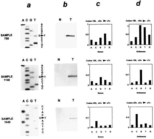
Figure 2.
Comparison of direct dideoxynucleotide sequencing (a), mutation-specific oligonucleotide hybridization (b), and the p53 GeneChip assay (c and d) in sequence analysis of the p53 gene. Relative fluorescent intensity is shown for the sense (c) and antisense (d) oligonucleotide probe sets of the GeneChip but not for the 14 additional probe sets present at each of these base pairs. Sample 758 contains a missense mutation (cgc → ctc at codon 158) detected by all three techniques. Sample 1,140 contains the same mutation as sample 758 present as a faint band on the sequencing gel. However, it was not detected by the p53 GeneChip assay. A mutation at codon 253 in sample 1,049 was detected by the p53 GeneChip assay and confirmed by mutation-specific oligonucleotide hybridization despite its absence by direct sequencing.

