Figure 2.
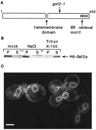
Localization of Gsf2p to the ER. (A) Schematic diagram of Gsf2p indicating the position of a putative transmembrane domain (residues 177–198, LVAQWLFFVMHIFKVGIITLFL), the gsf2-1 mutation (Q260 CAG codon → TAG stop codon), and a C-terminal dilysine (KKXX) ER-retrieval motif (10, 11). (B) Immunoblot analysis of HA–Gsf2p. A spheroplast lysate was prepared from a gsf2Δ strain (PS352) carrying pHA–GSF2 and was centrifuged at 13,000 × g. The P13 fraction was resuspended in buffer. Aliquots were washed with buffer (mock) or with buffer adjusted to 1 M NaCl or 1% Triton X-100 and centrifuged at 13,000 × g to generate pellet (P) and supernatant (S) fractions. Proteins were resolved by SDS/10% PAGE, and HA–Gsf2p was detected by immunoblotting with anti-HA antibody. (C) Fluorescence microscopy of GFP–Gsf2p. The gsf2Δ strain PS352 was transformed with pGFP–GSF2, and cultures were grown to mid-logarithmic phase in SC-Leu + 4% glucose. A 0.5-ml aliquot was centrifuged briefly at low speed, and 1 μl of a concentrated cell suspension was visualized by using fluorescence microscopy. Exposure time was 2 sec. (Bar = 2.5 μm.)

