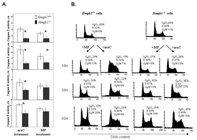
Fig. 1.

Hmgb1- deficient cells demonstrate decreased caspase activity and a compromised cell cycle arrest after genotoxic stress induced by antimetabolite drugs.
Panel A: Activation of caspases 2, 3, 8, and 9 in Hmgb1+/+ MEFs compared with Hmgb1−/− MEFs after 42 hr treatment with 10 μM MP or 0.5 μM araC for 42 hr. Activity of caspases was assayed using fluorogenic substrates as described in Materials and Methods. Relative activity of individual caspases in treated cells was expressed as a ratio to caspase activity in untreated cells after normalization per mg protein. Data are the mean ±SD generated from three independent experiments. ru, relative units. Asterisks denote significantly different levels of activity (p<0.05).
Panel B: Cell cycle arrest is induced by anticancer drugs in Hmgb1+/+ but not in Hmgb1−/−MEFs. Hmgb1+/+ (right) and Hmgb1−/− (left) MEFs were treated with 10 μM MP and 0.5 μM araC for 14 hr, 28 hr, and 42 hr, stained with propidium iodide, and DNA content was analyzed using flow cytometry after treatment with DNAse-free RNAse. The percentages of Hmgb1+/+ and Hmgb1−/− cells in G0/G1, S, and G2-M phases were determined by DNA histogram analysis.
