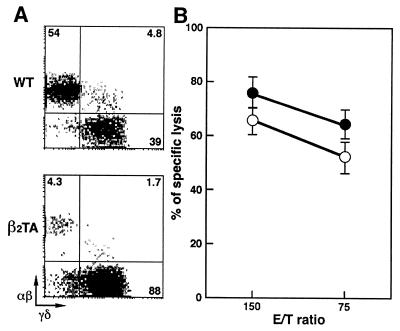
Figure 2.
Two-color flow cytometric analysis and cytolytic activity of IEL isolated from wt and β2TA mice at 2–3 months of age. (A) Representative staining of αβ- and γδ-IEL. IEL were incubated first with anti-TCR-αβ mAb (biotinylated) and then with streptavidin–phycoerythrin and anti-TCR-γδ mAb (FITC-conjugated). Percentage of positive cells in the corresponding quadrants is shown. (B) Redirected cytolytic activity of γδ-IEL from wt (○) and β2TA (●) mice in the presence of anti-TCR-γδ mAb. The results are means ± SD of data obtained from two independent analyses of four mice per group.

