
Figure 3. Pulse-chase study of hepcidin processing in HepG2 cells transfected with furin siRNA.
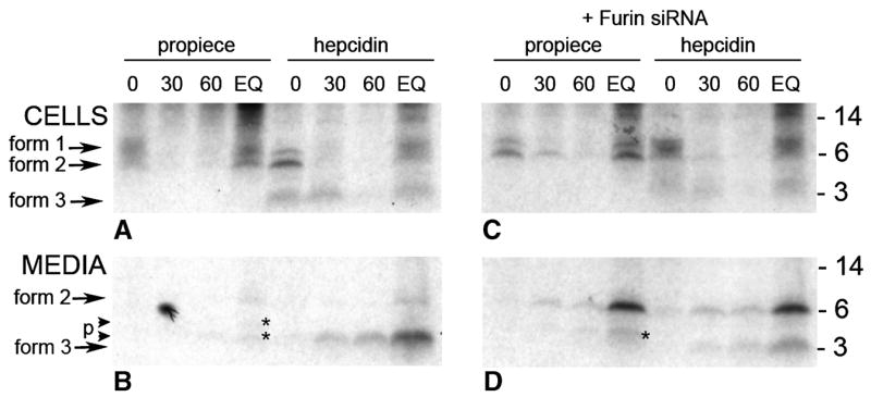
Cells were transfected twice over a 48 hr period with either blank vector (panels A and B) or vector containing furin siRNA (panels C and D). Cells were labeled with 35S-met-cys for 1 hr then subjected to cold chase for the times indicated. Lane EQ was labeled for 3 hr. Cell lysates (top panel) or the corresponding culture media (lower panel) were immunoprecipitated with either anti-pro (propiece) or anti-mature (hepcidin) antibody and analyzed on SDS-tricine PAGE. The autoradiogram of the gels are shown with the corresponding time of cold chase indicated above the lane (minutes). Molecular weight standards are indicated on the right panels. Three forms of hepcidin are seen as indicated by the arrows. Cleaved forms of the propiece are indicated by the small arrows (p) and the asterisks (panels B and D).