
Figure 1. Structure and subcellular localization of ROZA.
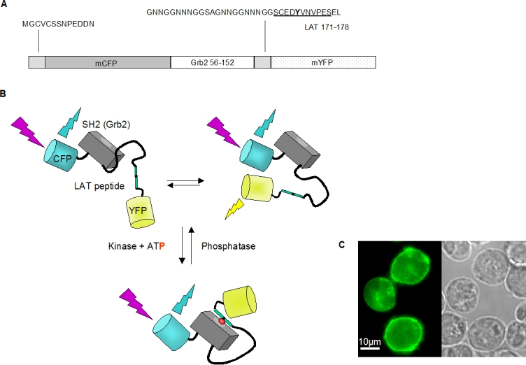
(A) Scheme of the ROZA sequence including the N-terminal residues from p56 Lck, the linker sequence and residues 171-178 of LAT (underlined) surrounding Y175 (in bold). (B) Model showing the schematic structure of ROZA (without its membrane anchor), and illustrating that, in the absence of ZAP-70 activity, ROZA may adopt several conformations, some of them allowing FRET. Following phosphorylation of the LAT based sequence by ZAP-70, ROZA adopts a constrained conformation incompatible with FRET. (C) In Jurkat T-cells transiently transfected with ROZA, the probe is mainly located at the plasma membrane as visualized by CFP fluorescence.