
Fig. 2.
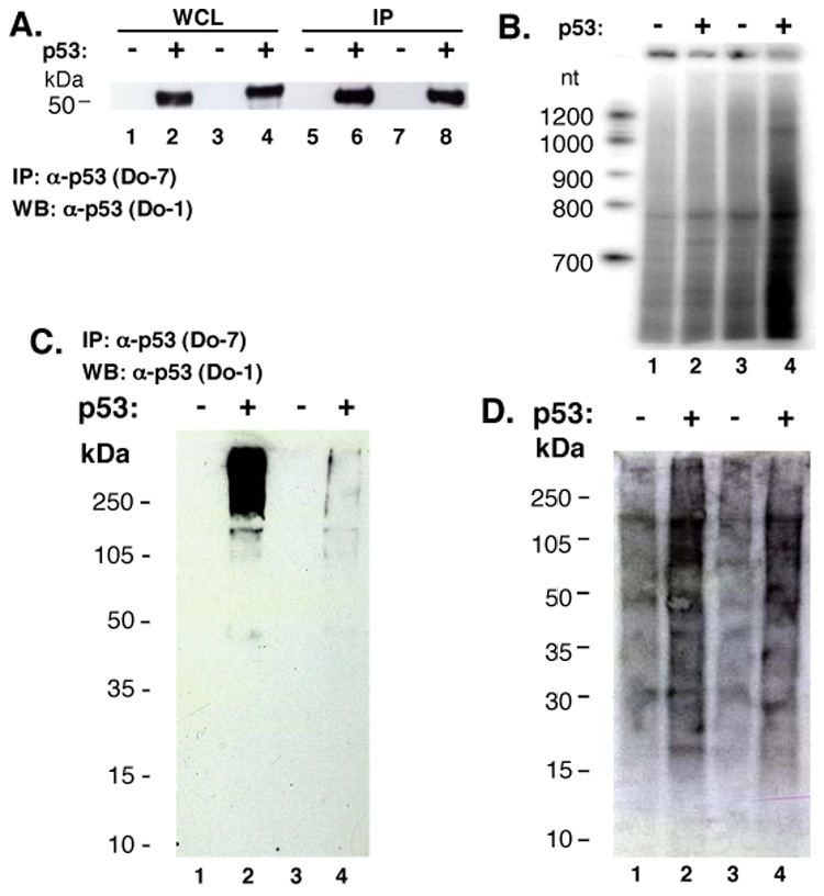
RNA is purified from cell lysates by anti-p53 antibodies regardless of p53 status. (A) Western analysis of p53. Preparations of whole cell lysate (WCL; 10% of total) [HCT116 (tp53 −/−) lane 1; HCT116 (TP53 +/+) lane 2; PC-3 (tp53 −/−) lane 3; MCF-7 (TP53 +/+) lane 4, after IP with anti-p53 Do-7 (lanes 5–8, respectively). (B) Co-immunoprecipitated RNA after [γ32P]-ATP labeling: HCT116 (tp53 −/−) lane 1; HCT116 (TP53 +/+) lane 2; PC-3 (tp53 −/−) lane 3; MCF-7 (TP53 +/+) lane 4. Formaldehyde cross-linked whole cell lysate: [HCT116 (tp53 −/−) lane 1; HCT116 (TP53 +/+) lane 2; PC-3 (tp53 −/−) lane 3; MCF-7 (TP53 +/+) lane 4], subjected to IP with anti-p53 Do-7 followed by RNA radiolabeling. (C) Western blot with anti-p53 antibody Do-1. (D) Membrane from panel (C) exposed to film.