
Fig. 4.
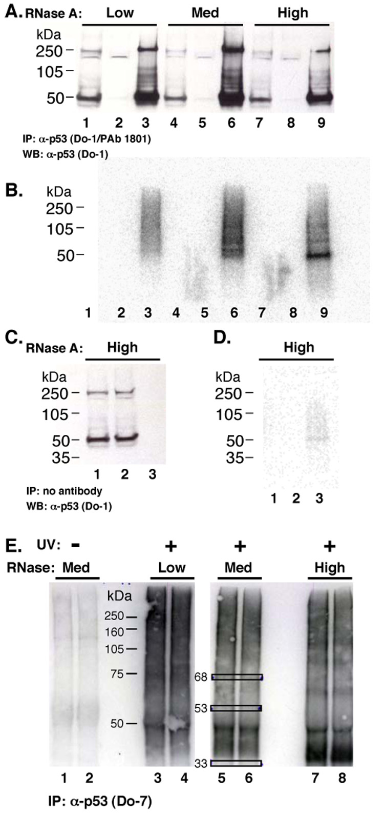
CLIP analysis of ribonucleoprotein “X”. (A) Western blot. MCF-7 cell lysate treated with RNase A. Lanes 1, 4 and 7: 10% of total WCL. Lanes 2, 5, and 8: cleared supernatant after IP with antibodies Do-1 and PAb 1801. Lanes 3, 6, and 9: IP material. (B) RNA analysis after p53 CLIP, RNAse treatment, and RNA labeling. Western blot from (A) exposed to a phosphorimager screen. (C) Mock IP without antibody, followed by processing, and Western blot. Whole cell lysate (lane 1), post-IP supernatant (lane 2) and IP sample (lane 3). (D) Phosphor image of membrane (C). (E) MCF-7 cell lysate treated with UV irradiation and RNase A, as indicated, subjected to IP by Do-7, and labeled. RNA samples of ~68, ~53, and ~33 kDa (boxes) were excised and cloned.