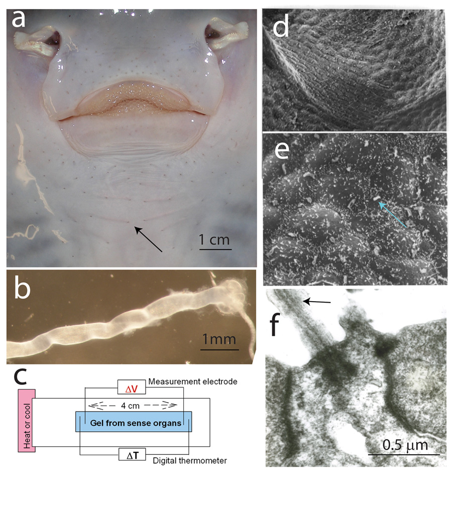
Fig. 1.

Ampullae of Lorenzini [9] are sense organs on the head of sharks [21], rays [5, 12], and chimaeras [10], containing a gel reported to have unique thermoelectric semiconductor properties [2]. (a) Visible as small pores around the oral surface of a skate (Raja erinacea) (arrow), the tubular organs, with an alveolus-shaped ending containing sense cells (b), are filled with an electrically conductive gel [24]. (c) Changes in voltage between two silver wire electrodes inserted in gel extracted from the organs were measured when a temperature gradient was imposed by heating or cooling one end of the apparatus. (d) Scanning electron microscopy of the sensory epithelium shows two types of cells: (e) sense cells, bearing a non-motile cilium (arrow), and mound-shaped support cells. (f) A cilium shown by transmission electron microscopy. (d–f) from Hydrolagus colliei. Previous studies show no ultrastructural differences in the sensory epithelium of chimaeras and elasmobranchs [11–14].
