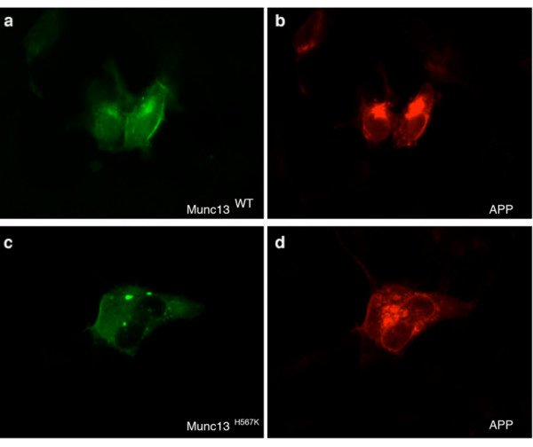
Figure 2.
Localization of APP following PMA treatment. HEK293 cells were co-transfected with the following cDNAs: (a, b) APPSWE-pPrk5 and Munc13-1WT-pEGFP-N1 and (c, d) APPSWE-pPrk5 and Munc13-1H567K-pEGFP-N1. Treatment with 100 nM PMA was carried out for 2 hours. GFP immunofluorescence allowed visualization of (a) Munc13-1WT and (c) Munc13-1H567K mutant molecules (green). (b, d) APP was immunolabeled with rabbit polyclonal anti-APP-specific antibody 369 followed by rhodamine red conjugated secondary antibody (red).

