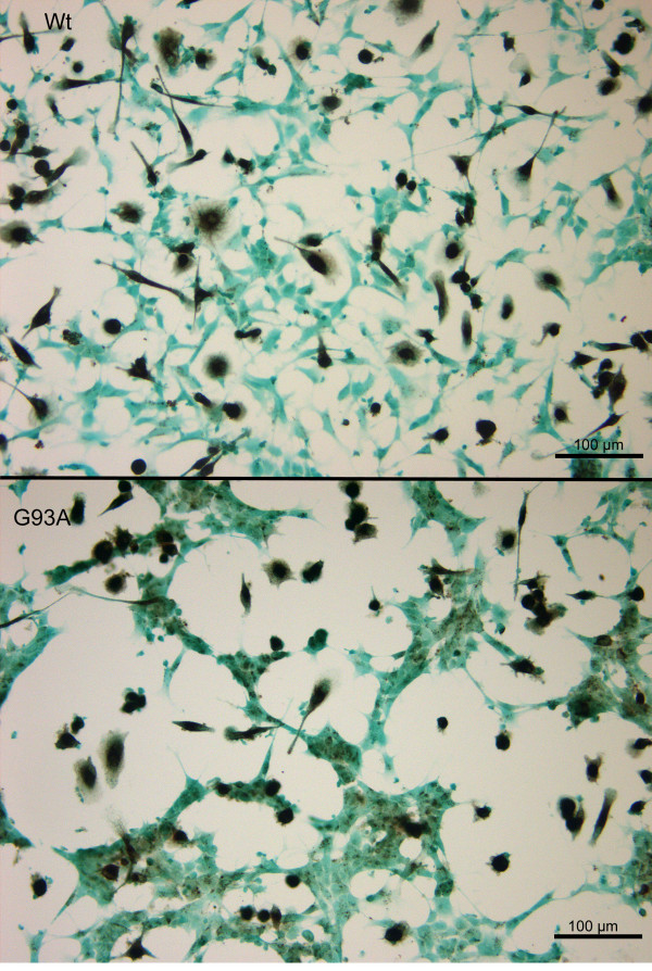
Figure 12.
Vulnerability of G93A-SOD1 and wild-type SOD1 neuroblastoma cells to the attack of monocytes stimulated with Pam3CSK4. After staining of Pam3CSK4-stimulated co-cultures with light green and macrophage staining with CD 68 less neuronal cell somata of G93A-SOD1 SH-SY5Y (lower panel) cells than of Wt-SOD1 cells in equally treated co-cultures (upper panel) were visible. Please note the clustering of severely damaged/dead G93A-SOD1 cells and the area devoid of neuronal cells in the vicinity of groups of macrophages.

