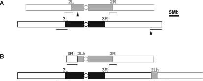
Figure 2. Schematic View of D. melanogaster Major Autosomes, Chromosomes 2 and 3.
(A) The chromosomes of a wild-type strain showing location of heterochromatin (filled rectangles), euchromatin (open rectangles) on left (L) and right (R) arms, centromeres (circle), and regions (underlined) that were included in the tiling microarray. The triangles indicate the positions of the breakpoints in the T(2;3)ltx13 translocation.
(B) Structure of the T(2;3)ltx13 translocation. Heterochromatin of 2L is denoted 2Lh.

